PATIENTENBIBLIOTHEK · NEUROCHIRURGIE
Diagnosing low back pain: from examination to MRI and specialist tests
When your lower back hurts, it is natural to want a scan that shows the exact cause. But assessment has a more complex purpose: first to rule out a serious condition, then to understand whether the pain involves a nerve, joints, muscles or another body system, and to choose the care you actually need.
No single test answers every question. MRI shows many structures clearly, but it does not measure pain. An examination assesses movement and nerve function, but cannot replace imaging when a serious condition is suspected. Good diagnosis brings together symptoms, examination findings, test results and changes over time.
This article covers the lower back and lumbosacral region. The list below is not a package of tests everyone needs. Some methods are used frequently, others only when a particular condition is suspected, and some invasive procedures remain controversial.
1. When assessment cannot wait
New inability to pass urine, loss of bladder control together with neurological symptoms, numbness around the genitals or anus, or rapidly worsening weakness in one or both legs require immediate in-person medical attention. These may signal serious damage to nerve structures. Do not wait for a routine MRI appointment or an online consultation. 7,8
Urgent medical assessment is also needed for pain after significant trauma, especially with osteoporosis; fever with a risk of infection; or unusual new symptoms in someone with cancer. Sudden severe back and abdominal pain with fainting or marked weakness requires emergency care: the cause may lie outside the spine.
An individual warning sign does not establish a diagnosis. The combination of symptoms, their progression and risk factors matter. However, when a dangerous condition is suspected, a doctor does not have to wait several weeks or complete a course of treatment before investigating. 7,9
Cauda equina syndrome involves the bundle of nerve roots in the lower spinal canal. All the “classic” signs do not have to be present at the same time. A normal result on one test cannot safely rule it out on its own.
2. Talking to your doctor: the first diagnostic step

Your doctor asks where the pain starts, whether it stays in the back or travels into the buttock and leg, when it began and how it changes. Numbness, weakness, changes in walking, bladder function or sensation around the genitals and anus, injuries, fever, previous operations and medicines are important.
Sleep, work, physical demands, other conditions and what has already helped also matter. Pain while standing, walking, sitting or at night helps form a working explanation, but cannot reliably identify a particular disc or joint on its own.
This conversation is called taking a medical history. It guides the next steps. A brief referral saying “low back pain” gives a radiologist much less information than a clear clinical question. 1,2
3. Examination, walking and movement

Your doctor observes how you stand up, walk and change position, and assesses posture, available movement and tender areas. When needed, they also check foot pulses, hip joints and other regions: not all pain near the lower back comes from the spine.
Feeling the tissues is called palpation. It helps locate tenderness but does not prove the precise source of symptoms. A tender muscle may be reacting to another problem, while prominent changes on a scan may cause little restriction of movement.
Pelvic position, uneven shoulders and pain on bending are parts of the overall picture. None alone proves that a vertebra is “out of place”, the pelvis is “misaligned”, or a disc necessarily needs surgery. 2,3
4. Neurological examination: how the nerves work
Testing muscle strength
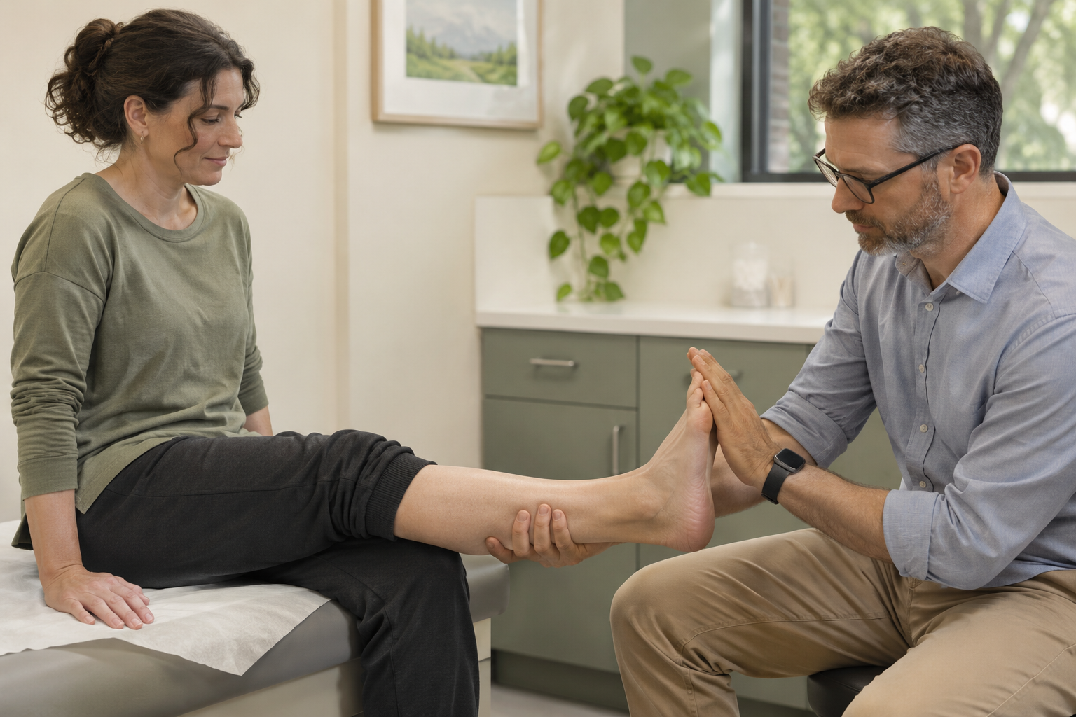
Your doctor asks you to perform movements and compares both sides: for example, lifting the foot, straightening the big toe or knee, or rising onto your toes. Gentle resistance may be added. It is important to distinguish true weakness from movement limited by pain.
The pattern of weakness in particular muscles helps suggest the level affected. However, muscles do not receive signals from just one isolated nerve root, so findings are interpreted alongside other signs. New or worsening weakness changes the urgency of assessment. 2,8
Testing sensation

Light touch and other safe tests are used to compare sensation in the legs. Your doctor asks about numbness, tingling, burning and unusual tenderness. The distribution helps distinguish a nerve root problem from damage to an individual peripheral nerve or a more widespread nerve condition.
Maps of skin sensation are approximate: areas supplied by different roots overlap and vary between people. An online “dermatome” diagram alone cannot reliably identify the affected root. 2,4
Testing reflexes
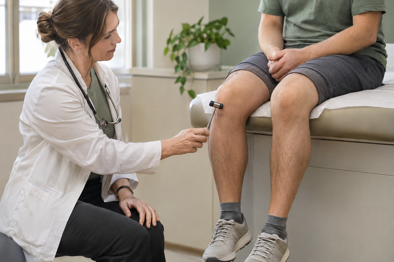
A reflex hammer is used to check knee, ankle and, when necessary, other reflexes. Differences between sides and the combination with strength and sensation matter. A reduced or absent reflex does not always indicate a new dangerous problem, and preserved reflexes do not exclude every type of nerve root disorder.
Radicular pain and radiculopathy are not exact synonyms. Radiculopathy means impaired nerve root function, which may cause weakness, altered sensation, changes in reflexes or electrophysiological abnormalities. Radicular pain can occur without an obvious motor deficit.
5. Specific clinical tests
Straight leg raise — the Lasègue test
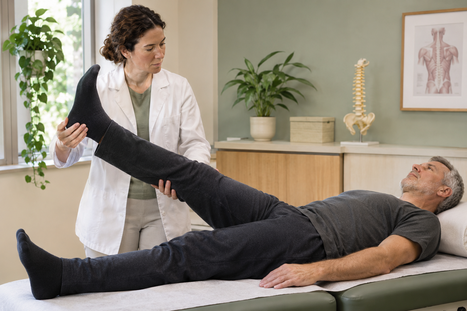
While you lie on your back, your leg is gently raised to assess whether familiar pain appears and where it travels. Reproduction of your usual leg pain is more relevant to a root problem than simply feeling a stretch in the back of the thigh.
Sometimes the crossed straight leg raise and other tests of nerve tissue sensitivity to movement are used. A positive test does not prove a disc herniation; a negative test does not exclude every cause of pain. Do not force your leg through severe pain to “check the diagnosis” yourself. 2,8
Hip and sacroiliac joint tests

Your doctor changes the position of the hip and gently loads certain structures, asking where and what kind of pain occurs. This helps identify a possible hip problem or involvement of the sacroiliac region.
One test cannot confirm the source with complete accuracy. For the sacroiliac complex, several tests are considered together and other causes are excluded. Pain can arise from surrounding ligaments as well as the joint space; this matters when interpreting a subsequent injection. 5,16
6. Questionnaires and everyday function

Pain scales and questionnaires such as the Oswestry Disability Index or Roland–Morris questionnaire help describe limitations in walking, sitting, sleeping, dressing and working. They are useful for comparing your condition before and after treatment. They do not identify which disc is painful.
Anxiety, mood, fear of movement and work circumstances may also be assessed. This identifies factors affecting recovery; it does not mean the pain is imaginary. Persistent, complex symptoms may benefit from assessment by several professionals working together. 7,10
7. When imaging is needed — and when it can wait
For a typical new episode without warning signs, early imaging often does not change initial care. The American College of Radiology, US Department of Veterans Affairs and Department of Defense guidelines, and the UK's National Institute for Health and Care Excellence do not recommend immediate imaging for every patient. 7,9,10
If pain persists despite appropriate treatment, substantially limits daily life, or an intervention is being considered, your doctor reassesses the need for imaging. Around six weeks is often discussed, but this is neither a universal ban on earlier imaging nor an automatic referral for everyone on day 42. 9,11
The key question is: which decision will the result change? Suspected nerve compression, fracture, infection, tumour or inflammatory disease requires a different pathway from nonspecific pain. German guidelines for specific low back pain also emphasise matching imaging to the clinical question. 12
Repeating an unchanged study simply because several months have passed is usually unnecessary. However, new symptoms, an injury, worsening weakness or a change in the suspected diagnosis may justify another investigation.
8. Magnetic resonance imaging — MRI
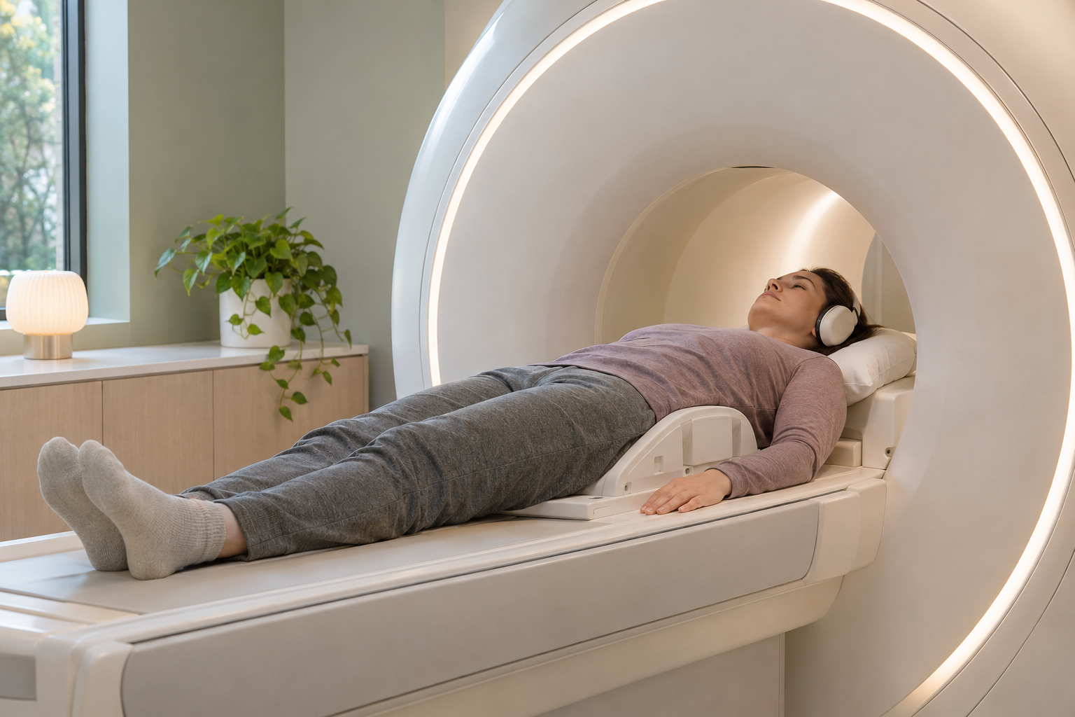
MRI uses magnetic fields and radio waves, without X-rays. It shows discs, nerve structures, ligaments, bone marrow and many soft-tissue changes well. It is often chosen for relevant nerve root symptoms, suspected spinal stenosis, infection, tumour or another condition that needs clarification. 1,3,9
You usually lie still on a table inside the scanner. The machine makes loud noises, so hearing protection is used. If pain or claustrophobia is a problem, tell the imaging department beforehand so arrangements can be discussed.
For a routine question about a herniated disc, MRI is often performed without contrast. Contrast may be needed for suspected infection, tumour or certain questions after surgery. It is not a compulsory “quality upgrade” for every patient. Report implants, electronic devices, metallic foreign bodies, pregnancy and kidney disease in advance; compatibility is checked for the specific device. 13
An MRI report describes anatomy. Disc protrusions, loss of disc height and other degenerative changes also occur in people without pain. What matters is whether the level, side and nature of the finding match the symptoms and examination. The word “herniation” alone does not determine the need for surgery. 26
If an inflammatory condition involving the sacroiliac joints is suspected, a separate examination of that region may be needed. A routine lumbar MRI does not answer every rheumatological question. 12,14
9. X-rays and movement views
Standard X-rays
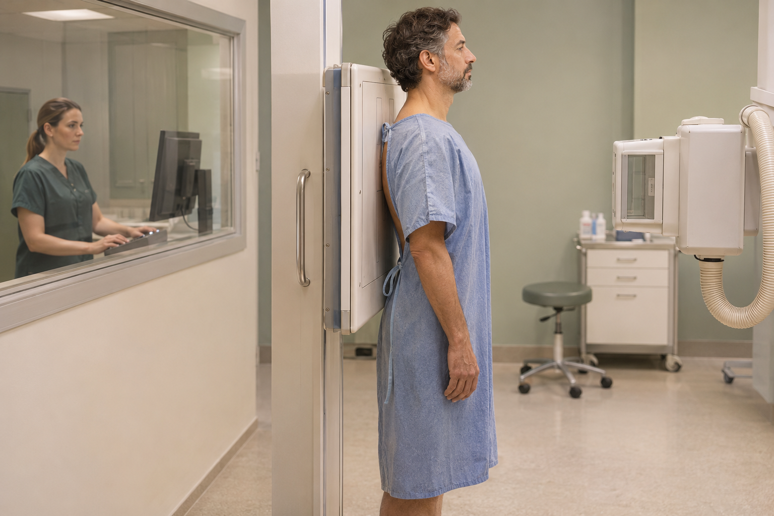
X-rays show bones, vertebral alignment, deformity and some signs of fracture. Standing images are sometimes needed to assess the spine under load. Discs and nerves are barely visible directly on standard X-rays: a narrowed disc space cannot prove the source of pain.
The method may help answer a specific question, but is not a compulsory step before every MRI. With dangerous neurological symptoms, the necessary investigation must not be delayed to follow a rigid “X-ray first” sequence. X-rays involve ionising radiation. 1,3,9,12
Flexion and extension views
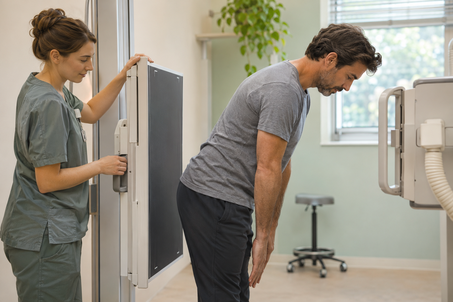
These are X-rays taken in different positions, sometimes used to investigate abnormal movement or instability. They are requested when the result may affect treatment or surgical planning, rather than for every episode of pain on bending.
Movements follow instructions and remain within a tolerable range. If a recent unstable injury is suspected, this is not a test to try independently. Normal movement views do not exclude every cause of pain, and any measured displacement must be interpreted in the overall clinical context. 2,12
10. Computed tomography — CT
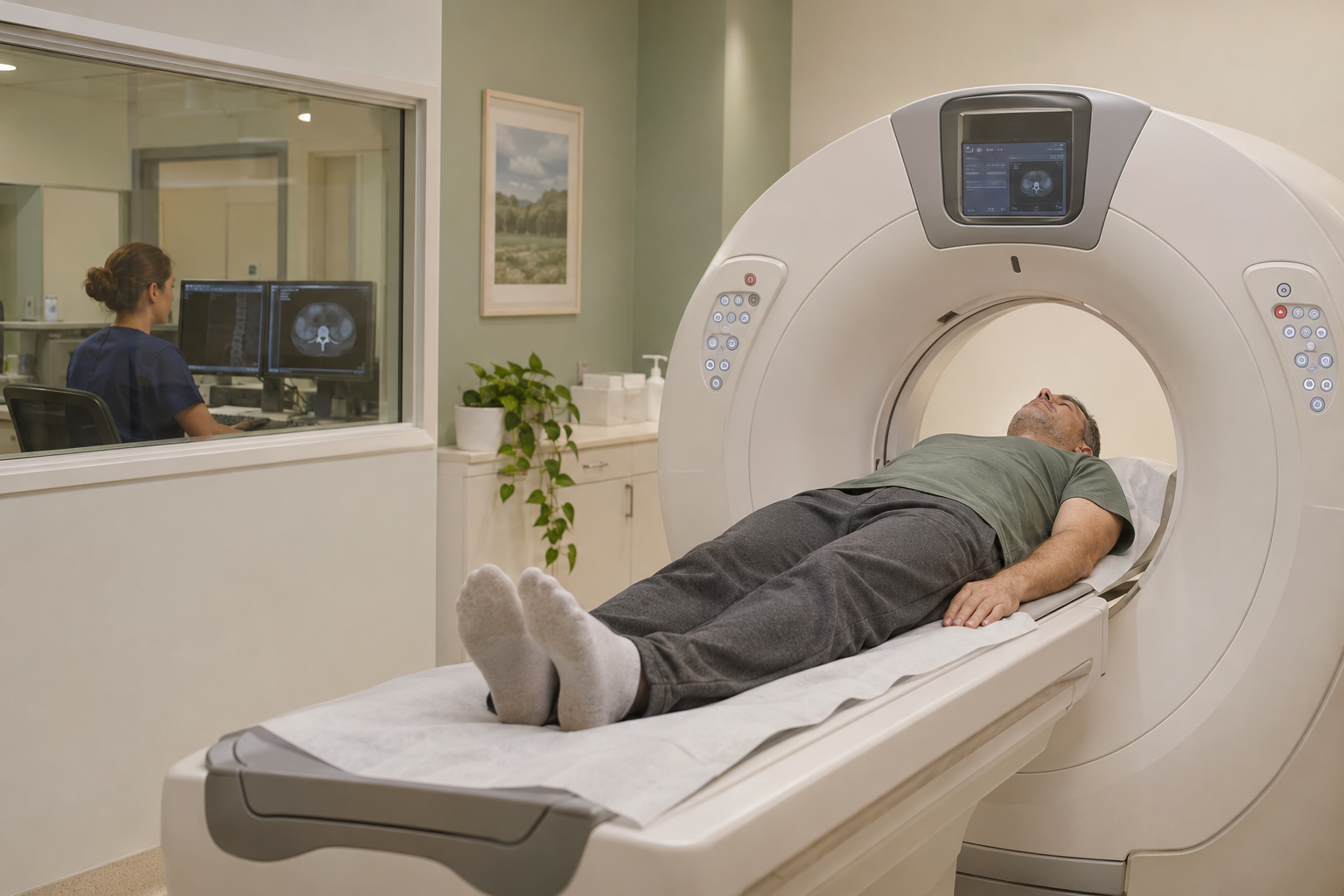
CT uses X-rays to produce detailed cross-sectional images. It is particularly useful for bone detail: complex fractures, certain bony defects, the position of metal implants and assessment of bone fusion after surgery. It may be chosen when MRI cannot be performed or does not answer a specific question.
MRI is usually more informative for nerves, discs and many soft-tissue changes. CT and MRI are therefore different examinations, and not everyone needs both. Whether contrast is needed depends on the question; many bone assessments do not require it. 1,2,9
11. Electrodiagnostic testing: nerve and muscle function
Needle electromyography — EMG
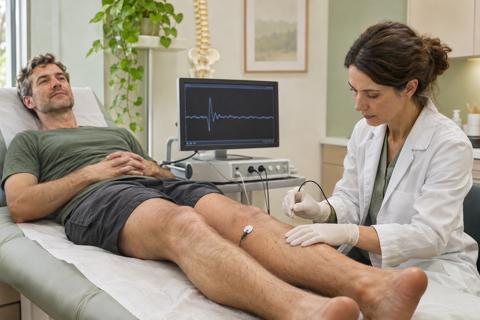
A thin electrode is inserted into selected muscles to record their electrical activity at rest and during contraction. No medicine is injected. The test helps assess signs of motor nerve fibre damage and distinguish some nerve root disorders from other neuromuscular conditions.
It is not needed for every episode of low back pain and does not show the size of a herniation. The examiner selects muscles according to the suspected level; briefly checking one randomly chosen muscle may not answer the question. You may feel a needle prick and some discomfort. Tell the examiner about medicines affecting blood clotting. 4,6,15
Nerve conduction studies
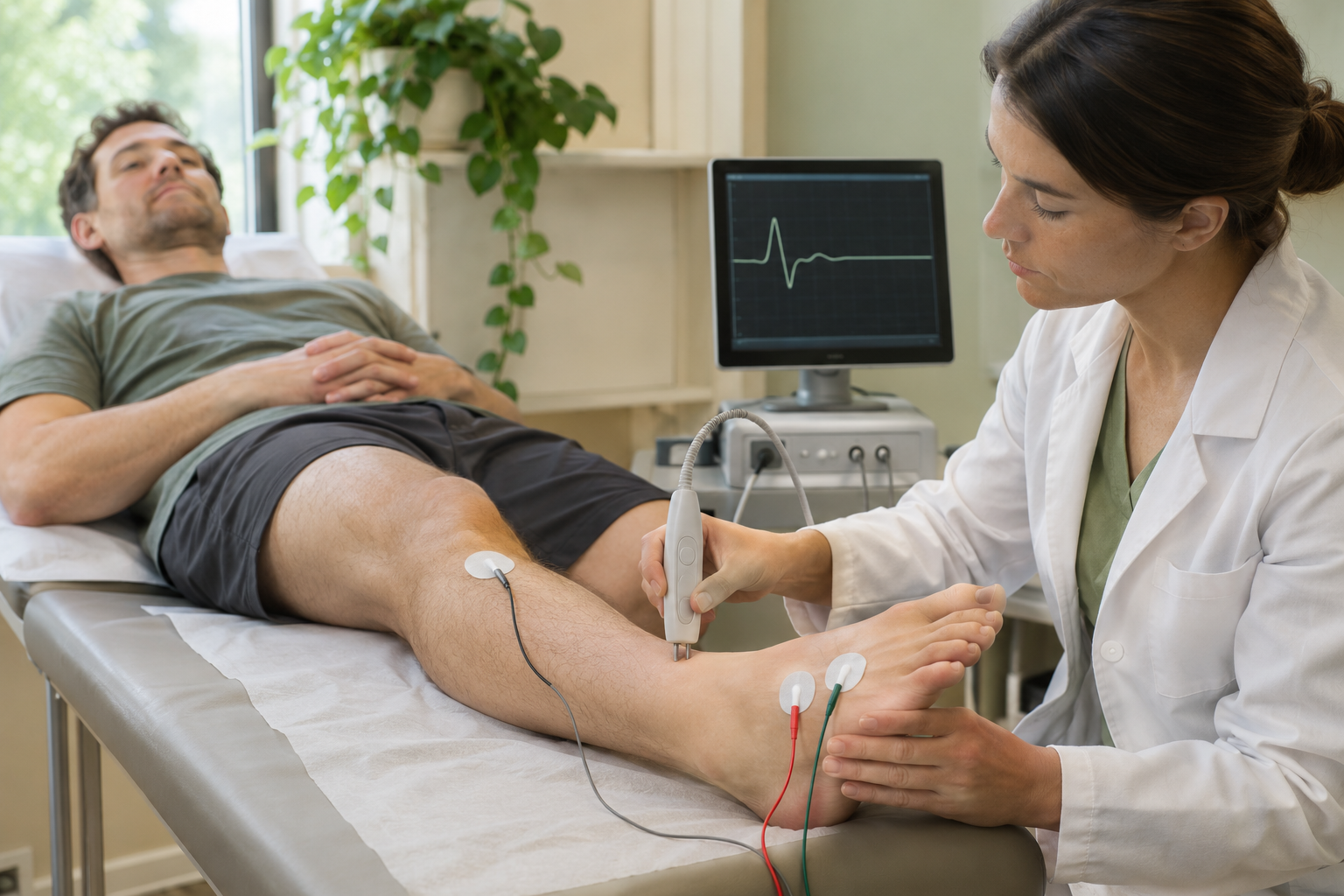
Brief electrical stimuli are applied through surface electrodes and the response is recorded. This helps identify a peripheral nerve injury or polyneuropathy, which may explain numbness and weakness differently from a spinal problem. This part is often combined with needle EMG. 15
Normal electrodiagnostic findings do not exclude all radicular pain. Changes may be absent when symptoms are mainly sensory or at an early stage; some abnormalities take time to appear. With worsening weakness or suspected cauda equina syndrome, these tests must not delay urgent imaging and care.
12. Blood tests and other laboratory investigations

There is no universal blood test for “the cause of low back pain”. Broad testing panels are usually unnecessary for typical nonspecific pain without warning signs. When a particular condition is suspected, a doctor may request a blood count, C-reactive protein, erythrocyte sedimentation rate or other tests.
Suspected infection sometimes requires blood cultures and further specialist tests. For inflammatory pain, a rheumatologist may request HLA-B27 testing. This is a marker of susceptibility, not a positive or negative “test for ankylosing spondylitis”: a positive result does not prove the condition and a negative result does not exclude it. Normal inflammatory markers also do not rule out all forms of spondyloarthritis. 14,17
Laboratory results are interpreted alongside symptoms and imaging. Tests needed before contrast administration or an invasive procedure may assess safety rather than identify the source of pain.
13. Other noninvasive investigations for selected situations
Ultrasound
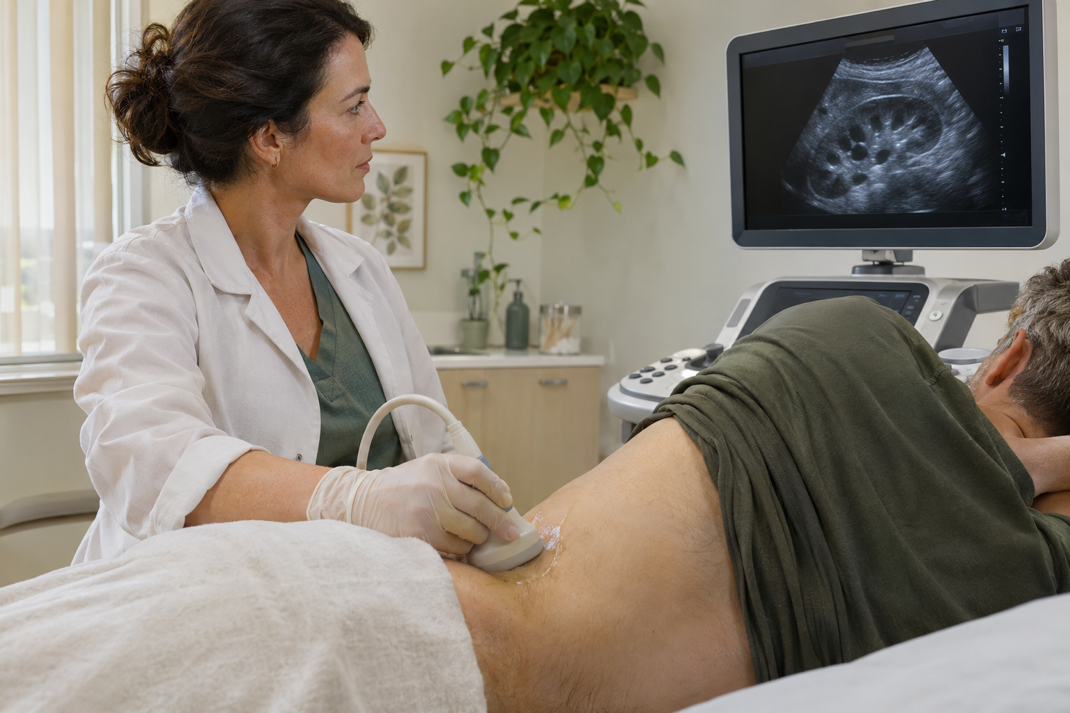
Ultrasound is useful when a doctor suspects a cause outside the spine, such as a kidney, abdominal organ or blood vessel condition. It can also measure urine remaining in the bladder after voiding. Such checks must not delay care when dangerous nerve damage is suspected.
Ultrasound does not replace MRI for routine assessment of lumbar discs and nerves inside the spinal canal. Using ultrasound to guide a needle is a separate application and does not mean that the cause of pain has been established. 1,8
Bone density measurement — DXA
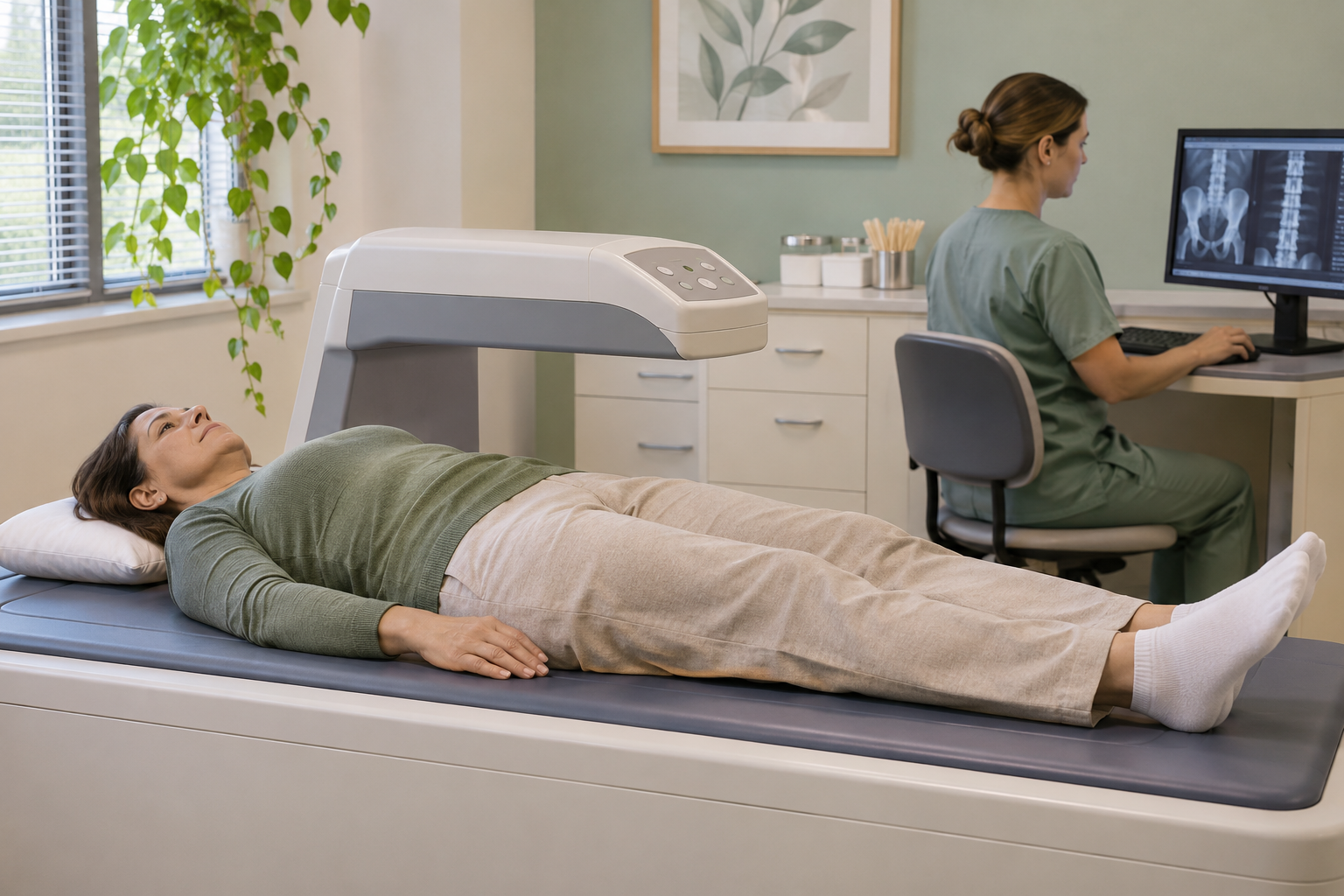
Bone densitometry, often called DXA, helps identify osteoporosis and assess fracture risk. The lumbar spine and hip region are usually examined. A small dose of X-rays is used.
It does not assess disc herniation or directly identify painful tissue. If a vertebral fracture is already suspected, appropriate imaging is needed to establish its presence and features. Arthritis, deformity, fractures and metal implants can affect lumbar measurements, so interpretation matters. 18
Bone scintigraphy and SPECT/CT
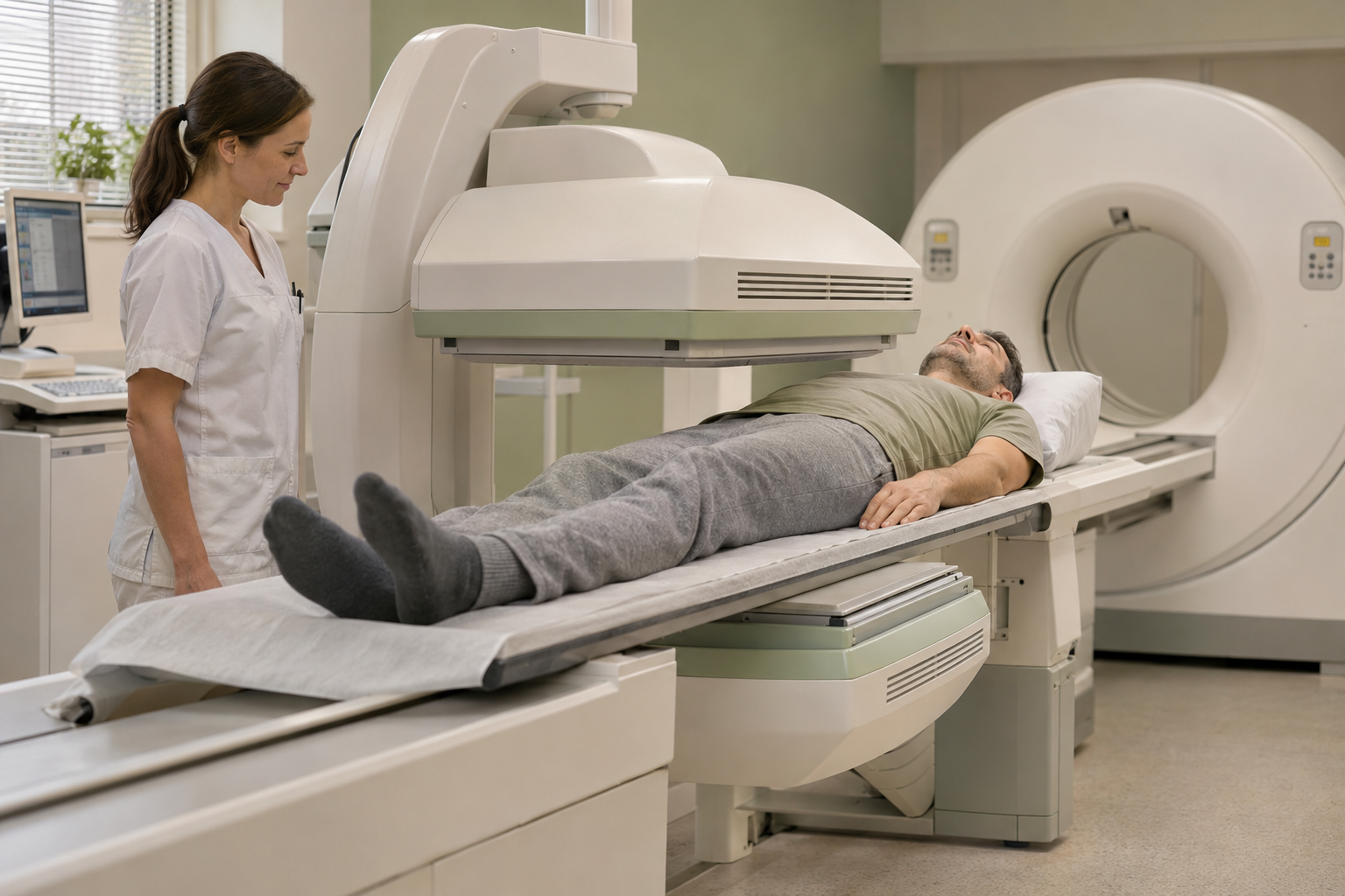
After a small amount of radiotracer is administered, a special camera records its distribution. The method reflects bone metabolic activity; combining it with CT helps locate a change more precisely.
This is a specialist investigation for selected questions, such as clarifying certain bone changes, an occult injury or postoperative findings. It is not the routine next step after every MRI. Increased uptake does not establish one specific diagnosis: different processes can cause it. There is radiation exposure; report pregnancy or breastfeeding in advance. Scintigraphy is not routinely recommended to investigate axial spondyloarthritis. 14,19
14. Invasive diagnostic procedures: a specific question is essential
These procedures are not a compulsory stage for someone with persistent pain. They are considered when the expected answer may change a decision and simpler methods have not provided the information needed. Risks, alternatives and interpretation of the result should be discussed beforehand.
CT myelography
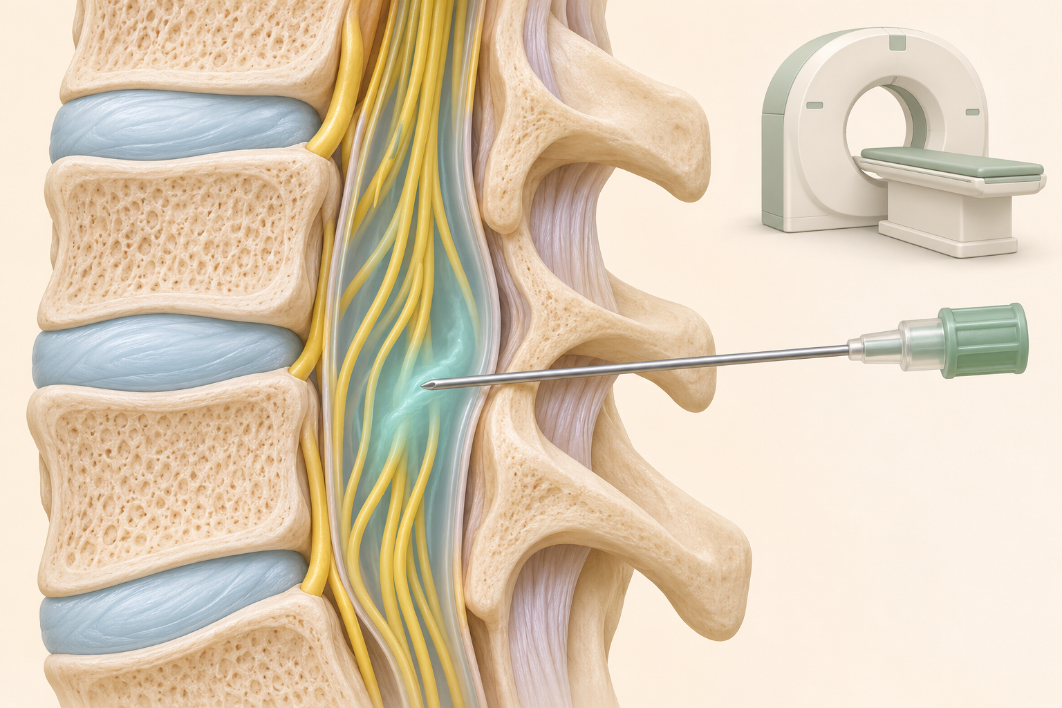
Contrast is introduced through a lumbar puncture into the space containing cerebrospinal fluid, followed by imaging, often CT. This differs from an epidural injection: the diagnostic contrast is inside the covering of the nerves, rather than outside it.
The investigation may be needed if MRI is contraindicated, severely affected by metal artefact or leaves an important question about nerve structures unresolved. It involves a puncture, radiation and contrast. Complications can include post-puncture headache, infection, bleeding and other problems. It is not an upgraded replacement for MRI for everyone. 2,20
Medial branch blocks to assess joint-related pain
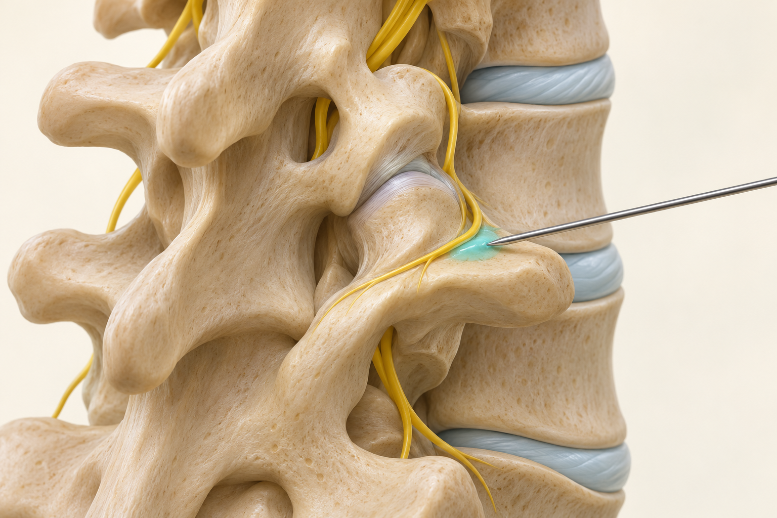
A small amount of local anaesthetic is injected near nerve branches that carry signals from the facet joints. The aim is to assess a change in familiar pain, for example before considering radiofrequency denervation. The injection is not into the joint itself or a major nerve travelling into the leg.
Several branches are usually relevant to one joint; the diagram shows one illustrative target. The clinician decides in advance which movements and sensations to compare, and when. Guidelines differ on the required degree of relief and whether repeat testing is needed. One positive response guarantees neither an exact diagnosis nor treatment success. 5,21
Diagnostic sacroiliac joint injection
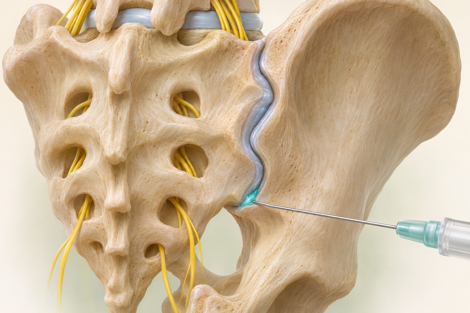
Local anaesthetic is injected into the joint under imaging guidance, and the change in familiar pain is assessed. This test is used in selected patients after clinical evaluation. Buttock pain alone is not a sufficient reason to perform it.
A positive response supports involvement of structures inside the joint, but does not fully assess surrounding ligaments or every cause of pain in this region. Spread outside the target can cause a false-positive result. No response also needs interpretation in light of the technique and initial hypothesis. 16
Selective diagnostic nerve root block
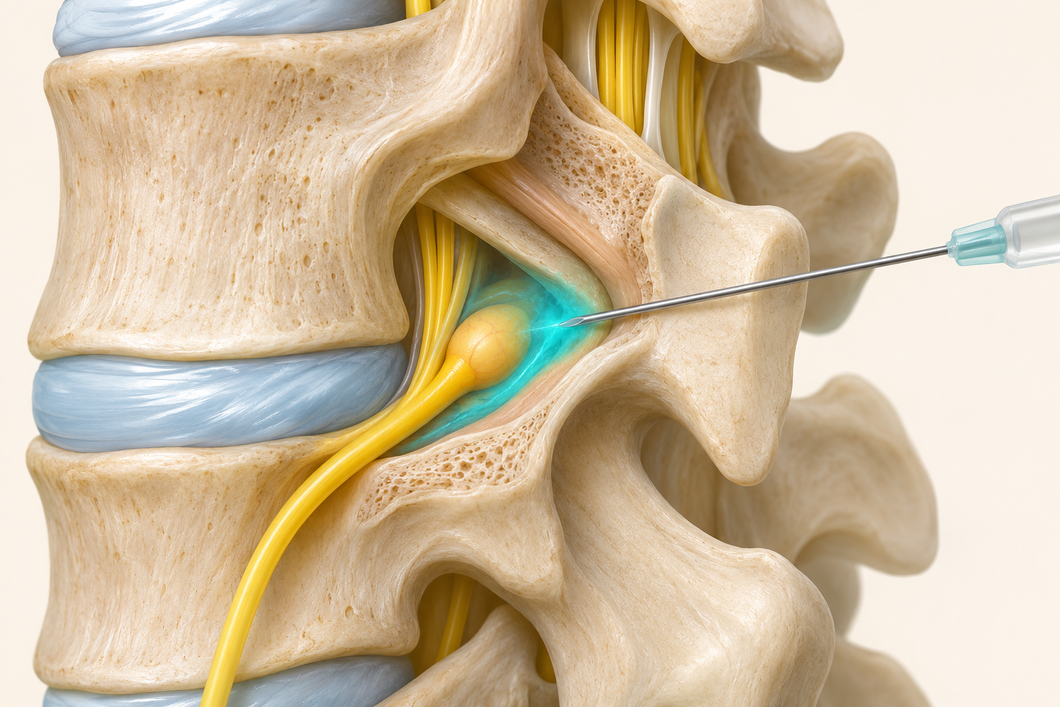
This is sometimes considered when symptoms and imaging do not clearly distinguish between several suspected roots. Anaesthetic is delivered near a specific area and temporary pain relief is assessed. It is not injected into the nerve itself.
The test is not an infallible way to select a level for surgery. Diagnostic accuracy is limited; solution spread and false-positive responses are possible. The result cannot be used independently of examination and imaging. A therapeutic injection intended to provide longer anti-inflammatory relief and a strictly diagnostic test have different purposes. 22
Provocative discography
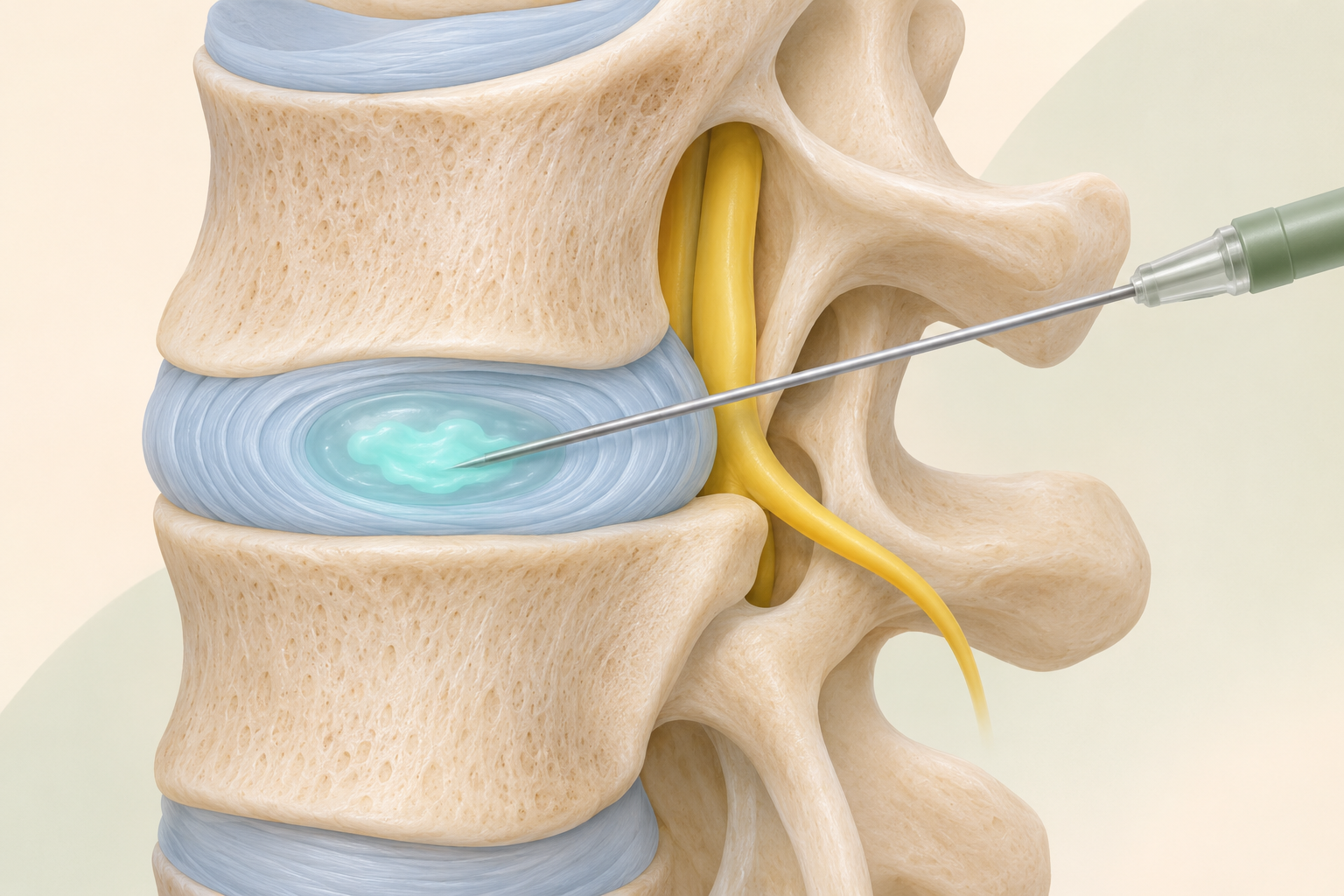
Contrast is injected directly into a disc to assess its structure and whether familiar pain is reproduced. Historically, this method was used to try to confirm a disc-related source before selected interventions.
However, discography is not a routine investigation for low back pain. Reproducing pain does not automatically prove its sole cause, and false-positive responses are possible. Risks include infection and disc injury. Current international recommendations do not consider its diagnostic value convincingly established for broad use. If it is proposed, ask which decision it would change and why it is considered necessary. 5,8,23
Vertebral or disc biopsy
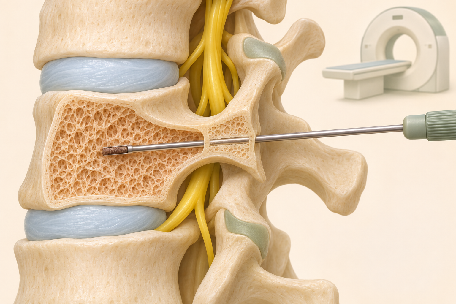
A biopsy takes a small tissue sample for analysis. It may be considered for a suspected tumour or infection when the nature of a change or the organism responsible needs to be identified. It is not a test for finding the cause of ordinary nonspecific pain.
The target and access route depend on the individual finding. Samples can be examined for tumour cells, inflammation and microorganisms. Sometimes the result is inconclusive and further assessment is needed. Risks depend on the route and the patient's condition, and include bleeding and infection. 17,24
Diagnostic blocks depend not only on pain reduction, but also on accurate targeting, anaesthetic spread, other medicines, expectations and changes in usual activity. A note saying “the block helped”, without the medicine, timing and response, provides less information than structured observation.
15. Bringing the findings together
A useful assessment answers both “what is visible?” and “what does it mean for this person?”. Do the side and level match the symptoms? Is nerve function impaired? Does the finding explain limited walking, or should another cause be investigated?
Several MRI findings do not mean several operations are needed. A normal MRI does not make real pain imaginary: scans cannot show every pain mechanism. Sometimes a sound assessment rules out a serious cause and establishes a follow-up plan without naming one exact painful tissue.
The working diagnosis is reconsidered when symptoms change. New neurological deficits matter more than simply comparing the millimetres in old and new disc reports. 2,7,9
16. Preparing for assessment and the role of an online consultation
Write down when symptoms began, how pain spreads, and any numbness, weakness or changes in walking. Prepare a list of medicines, conditions, operations and the effects of previous treatment. For image review, the original scan files are more useful than only a written report or a photograph of a screen.
An online consultation can help discuss whether an investigation answers the clinical question, whether another scan is actually needed and which assessments should be completed in person. A useful question is: “How will this result change my plan?”
An online conversation cannot replace a necessary neurological examination and is not a reason to wait with urgent symptoms. Agree preparation for MRI, contrast, puncture or a block with the department performing it. Do not stop anticoagulants or other regular medicines on your own.
17. Frequently asked questions
Why was I not offered MRI straight away?
Without warning signs, an early scan often does not change care. This does not mean your symptoms are being ignored. An examination, a clear plan and a review date matter; new symptoms can change the decision.
Which is better — MRI or CT?
It depends on the question. MRI is better suited to many nerve and soft-tissue questions; CT provides detailed bone assessment and helps with certain postoperative findings. Both are not automatically needed.
Does finding a herniated disc prove the pain source?
No. The finding must match the level, side and nature of symptoms and the examination. Similar changes can occur in someone without symptoms.
Do normal electrodiagnostic tests rule out a pinched nerve?
Not always. Results depend on the type of damage, time since symptoms began, and nerves and muscles tested. With worsening weakness, an old normal test cannot replace a new examination.
If a diagnostic block helped, will surgery definitely help?
No. Temporary relief provides additional information but does not guarantee the outcome of surgery or denervation. The accuracy of the procedure and the full clinical picture must be considered.
Should MRI be repeated every year for chronic pain?
Not automatically. Another study is requested when the clinical situation changes or a new question could affect treatment. The passage of time alone is insufficient.