Patient Library
Lumbar disc herniation
Why pain can travel down the leg, when you may need an MRI and when to discuss surgery.
A herniated disc can cause pain that travels from the buttock down the leg, as well as numbness or weakness. For many people, symptoms gradually ease over several weeks. During the examination, your doctor checks how well the nerve is working and whether the herniated disc explains your symptoms.
About disc herniation

Intervertebral discs sit between the bones of the spine. Each disc has a tough outer ring and a softer centre. A disc herniation occurs when some of the disc tissue moves beyond its usual boundary. Pressure on a nearby nerve root and inflammation around it can cause pain.[3] [4]
What does it feel like?
A common symptom is shooting or burning pain in one leg. It can run from the buttock to the lower leg and foot and may worsen when you cough or sneeze. Some people also notice tingling, numbness or weakness. For example, it can become difficult to lift the foot or stand on tiptoe. Pain confined to the lower back has many possible causes, so your doctor considers the full picture.[4]
Medical note. Pain that travels along an irritated nerve root is called radicular pain. Radiculopathy means that the nerve root is not working normally. This can cause weakness, reduced sensation or changes in reflexes.
Can a herniated disc shrink on its own?
Yes. Over time, the body may absorb part or all of the displaced disc tissue. Pain often eases as the inflammation settles. Your doctor assesses progress by checking how you move, what you feel in your leg and whether strength is returning.[2] [5]
Diagnosis
What does the doctor check?
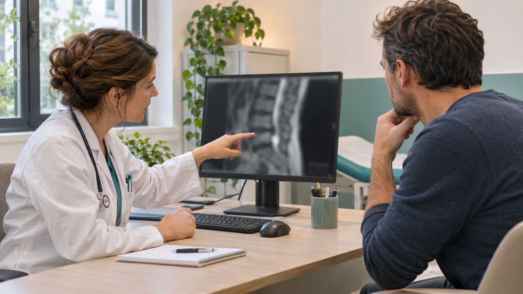
Your doctor asks where the pain travels, where you feel numb and whether your walking has changed. They compare strength, sensation and reflexes in both legs. They may ask you to walk on your heels and toes or gently raise your straightened leg to see whether this brings on your usual pain.[4]
When is imaging needed?
In the first few weeks, imaging can often wait if symptoms are improving and the examination shows no warning signs. Your doctor orders a scan when the result could change treatment, for example if leg pain persists and an injection or surgery is being considered. Marked or worsening weakness calls for earlier assessment. Suspected cauda equina syndrome requires emergency MRI.[1] [6] [7]
MRI – the disc, the herniation and nerve roots
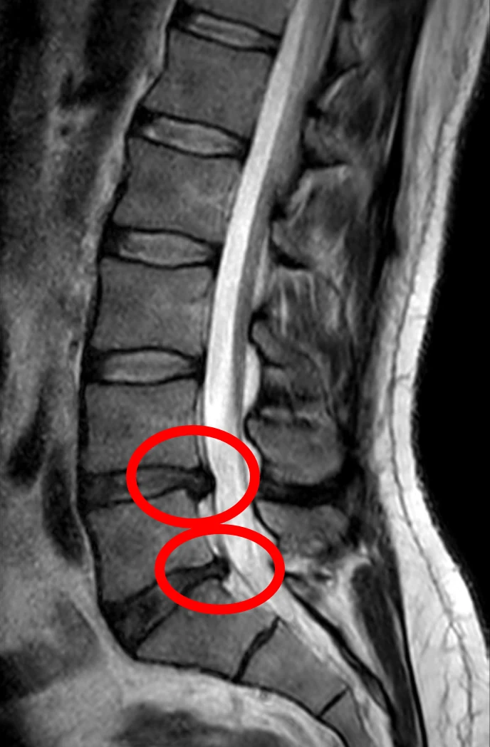
When a lumbar disc herniation needs to be confirmed, MRI without contrast is usually the preferred test. Side-view images show the disc levels, spinal canal and openings where the nerves exit. Cross-sectional images help locate the displaced tissue and show which nerve root it touches. MRI also helps assess whether the space around the nerve has narrowed.[4] [6]

CT – bone changes and the openings where nerves exit
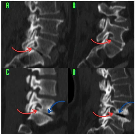
CT shows the bony edges of the vertebrae, joints, fractures and bone spurs in detail. Slices through a nerve exit opening can show whether a spur narrows it. CT can also show some disc herniations, although MRI usually gives a clearer view of soft tissues and nerve roots. Your doctor may choose CT to assess bone changes or when MRI cannot be performed.[6] [14]
X-ray – vertebral alignment and disc space height
A side-view X-ray can show how the vertebrae line up, how far apart neighbouring vertebral bodies are and whether there are large bone spurs. A smaller gap may indicate loss of disc height. An X-ray may help if your doctor suspects a fracture or a slipped vertebra. A plain X-ray cannot confirm a herniation or reliably show whether a nerve root is compressed in its exit opening.[6]
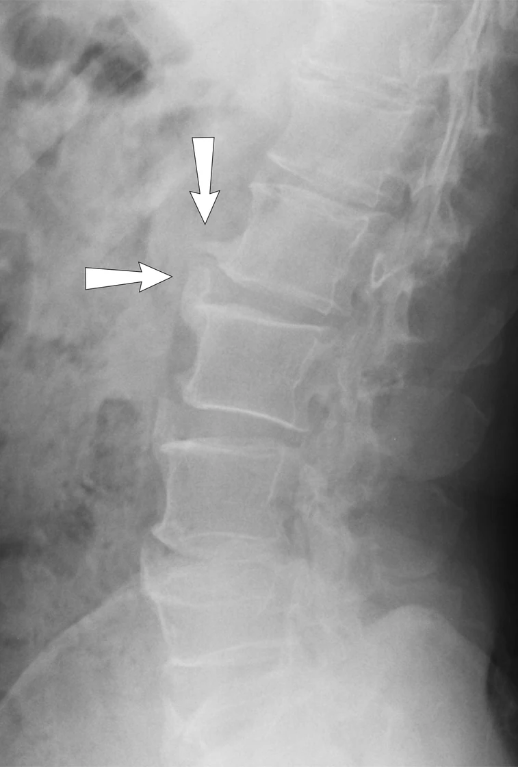

Medical note. The disc space lies between two vertebral bodies. The intervertebral foramen is the opening on the side through which a nerve root exits. Loss of disc height, bone spurs or a herniation can reduce the space available to the nerve. Narrowing of this opening is called foraminal stenosis.[14]
Disc protrusions and other disc changes are also seen in people without pain. Your doctor checks whether the side and location of the herniation match your symptoms and examination findings. The size in millimetres alone does not determine whether surgery is needed.[4] [8]
Medical note. Protrusion and extrusion describe the shape of displaced disc tissue. A sequestrated fragment has completely separated from the disc. These terms describe the scan findings. Treatment decisions depend on how the nerve is working and how much the symptoms affect daily life.[3]
Treatment

Treatment without surgery
If leg strength is preserved and there are no warning signs, treatment usually starts without surgery. At first, you can cut back on activities that sharply increase the pain and keep moving as tolerated, for example by taking short walks. Prolonged bed rest is usually unnecessary. As symptoms improve, a physiotherapist can help you choose exercises and gradually return to your usual activities.[7] [9]
Your doctor may suggest a short course of an anti-inflammatory painkiller if it is safe for you, taking into account stomach, kidney or heart conditions and any other medicines you take. It may provide limited relief for pain that travels down the leg. At follow-up, your doctor checks both pain and leg strength. If symptoms worsen, you need an earlier review.[7] [9]
When might an injection help?
If leg pain is severe and your doctor has identified the nerve root involved, you can discuss a targeted injection guided by imaging. This can temporarily reduce pain and make movement easier. The benefit varies. If weakness is worsening, urgent assessment of nerve function takes priority.[10] [11]
Medical note. Periradicular therapy (PRT) is a targeted injection into the epidural space through the opening where the nerve root exits. It is also called a transforaminal epidural injection.[10]
When is surgery considered?
Planned surgery may be considered if severe leg pain still interferes with walking, sleep or work after a course of treatment without surgery and the MRI findings match the symptoms and examination. The decision is often reviewed after about 6–12 weeks. Marked or worsening weakness calls for an earlier decision. Confirmed cauda equina syndrome caused by a herniated disc requires emergency assessment so pressure on the nerves can be relieved.[1] [2] [12]
In a microdiscectomy, the surgeon removes the disc tissue pressing on the nerve root. An endoscopic approach may be suitable in some cases. Spinal fusion is usually unnecessary for a first operation on an isolated disc herniation. In suitable patients, surgery can relieve leg pain sooner. On average, the difference compared with treatment without surgery narrows over time. Lower back pain, numbness and weakness can persist. Before surgery, your doctor discusses the expected benefits and risks, including a recurrent herniation, infection and a tear in the covering around the nerves.[12] [13]
Scientific references
Textbooks: AO Spine Textbook (2020), ch. 36, Surgery of the Lumbar Spine (2026), chs. 12 and 25, Handbook of Spine Surgery (2025), ch. 52.